
Every cell in your body is wrapped in a membrane about 4 nanometres thick. That's roughly 20,000 times thinner than a human hair. It's also very good at its job: it keeps water, ions and most medicines on the outside unless the cell lets them in.
A short, strong electric pulse can change that. This technique is called electroporation. The pulse opens tiny holes in the membrane, and molecules that couldn't get in before can now pass through. Researchers use it every day to get DNA into cells. Doctors use it to help chemotherapy drugs get into tumour cells, which is known as electrochemotherapy.
Opening the membrane is only half of what matters, though. A cell whose membrane doesn't close again is in trouble. So we asked two questions:
- How strong does the field have to be before the membrane opens?
- What happens once the field is switched off?
You can't film this under a microscope. The holes are a few nanometres wide and form in billionths of a second. So we built the membrane atom by atom and simulated it on eight GPUs using AxonOS.
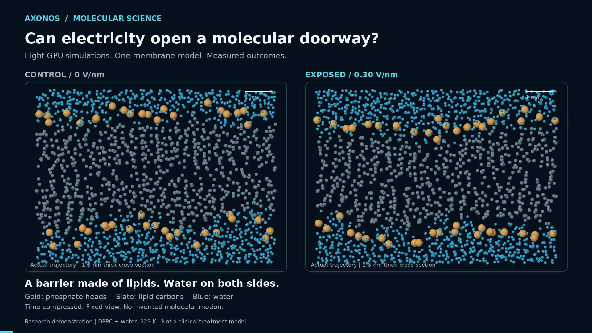
Left: a membrane with no electric field. Right: the same membrane after 26 nanoseconds under an applied field. Blue dots are water, grey are the oily lipid tails, gold are the lipid head groups. Every dot is a real atom position from the simulation.
The experiment in plain terms
We started from a published, well-studied model membrane (Ollila & Miettinen, Zenodo). It's made of DPPC, a common lipid often used as a stand-in for real cell membranes. Each lipid has two parts:
- a head that likes water (the gold dots in our images), and
- two long, oily tails that avoid water (the grey dots).
In water, the lipids arrange themselves into two layers, heads out and tails in. The result is a greasy sheet that water can't easily cross.

Before any field is applied, both membranes look the same: two rows of gold head groups with a dry, oily core between them and water on either side.
Our simulated patch is about 9 nm across. It contains 288 lipids and 8,756 water molecules, 63,708 atoms in total. We used GROMACS, a standard molecular dynamics engine. It calculates the forces on every atom and moves each one forward in time, 2 femtoseconds (two millionths of a billionth of a second) at a time, at 50 °C (323 K).
We then ran four conditions, each twice, with different random starting velocities:
Field strength | What it represents |
|---|---|
0 V/nm | Control: no field at all |
0.15 V/nm | Weak field |
0.30 V/nm | Moderate field |
0.50 V/nm | Strong field |
Each of the eight simulations followed the same three steps:
- Settle for 0.5 ns with no field.
- Field on for 30 ns, applied straight across the membrane.
- Field off for 15 ns, to see whether the membrane recovers.
That's 45.5 ns per simulation and 22.75 million time steps. Across all eight runs, we simulated 364 ns of molecular motion.
A note on the numbers: Simulations use stronger fields than a real cell would experience, so that effects that take microseconds in the lab show up within nanoseconds of simulated time. Treat these field values as settings inside the model. They are not doses and don't translate directly to a clinical device.
How we detected an opening
Pictures are persuasive, but we wanted a measurement. Our analysis script ran on the GPU and looked at a snapshot of the membrane every 20 picoseconds, about 2,250 snapshots per run. For each snapshot it did two things:
- Counted water molecules in the core. This is the number of water molecules within 1 nm of the membrane's centre. A healthy membrane has only a handful there.
- Checked for a connected water path. It linked together water molecules that sit closer than 0.35 nm to each other, which is about the distance between neighbouring molecules in liquid water. If a single connected chain of water stretched all the way through the oily core, from one side to the other, it counted as a core-spanning water pathway. This is the molecular sign of a pore. To count as a real opening, the pathway had to be present in three snapshots in a row.
To check that the result didn't depend on that 0.35 nm cutoff, we reran the analysis at 0.32 nm and 0.38 nm. The conclusions were the same.
What we saw
Weak fields: nothing happened
At 0 and 0.15 V/nm, the membrane held in both repeats. A few water molecules occasionally drifted into the core, typically around 10 to 20 at a time. This is normal thermal jiggling. No connected pathway ever formed, either during the 30 ns with the field on or during the 15 ns after.
Stronger fields: the membrane gave way
At 0.30 and 0.50 V/nm, all four simulations developed a pore. The stronger field opened the membrane much sooner:
Field | Repeat 1 | Repeat 2 |
|---|---|---|
0.30 V/nm | pore at 10.40 ns | pore at 6.52 ns |
0.50 V/nm | pore at 1.26 ns | pore at 1.20 ns |
The frame below comes from the video, just after the pathway appears in one of the 0.30 V/nm runs:
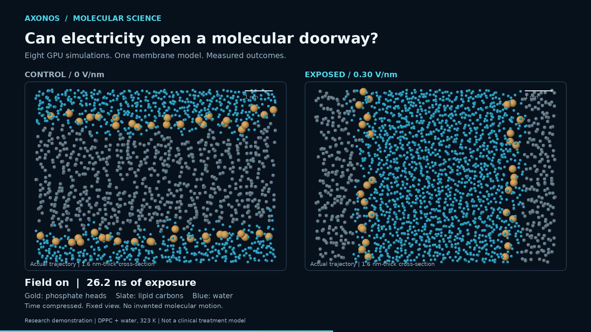
At 11 ns, a channel of water pushes through the membrane core. The gold head groups have followed the water inward and now line the walls of the pore.
Look at the gold dots. The water-loving head groups don't stay in their neat rows. They bend inward and line the walls of the new channel, so the water inside is surrounded by other water-loving molecules rather than by oily tails. Earlier simulation studies describe the same mechanism: water pokes a thin finger into the membrane, and the lipids rearrange around it to form a stable, water-lined pore.
The pores didn't stay small. The number of water molecules in the membrane core went from about 10 to roughly 2,400, more than a hundredfold increase:
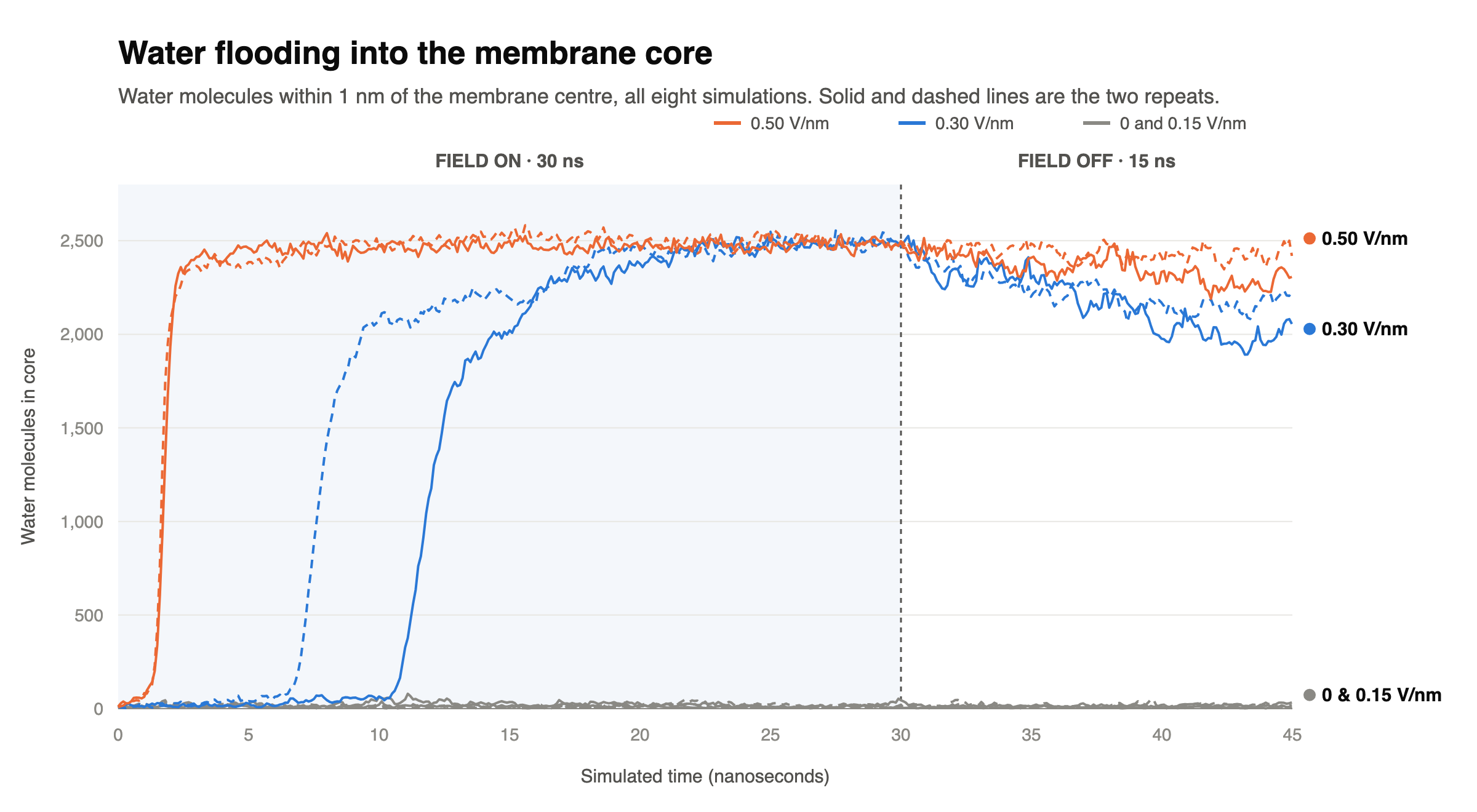
The main result in one chart. Each line is one simulation. The shaded region is when the field was on.
Field off: the door stayed open
At 30 ns we switched the field off and kept watching for another 15 ns.
None of the four pores closed. A connected water pathway was present in every single snapshot of the field-off period, including the very last one, in all four high-field runs. The amount of water in the core eased back a little, down about 5 to 17% by the end, but nothing close to resealing.

This doesn't mean the pores would stay open forever. Real membranes do reseal, over times far longer than 15 ns. What it does tell us is that once these pores formed, they didn't close quickly on their own. Opening a membrane and closing it are separate problems, and any useful electroporation protocol has to solve both.
What this study does and doesn't show
We think it's important to be clear about the scope of this result.
It shows that in this membrane model, fields of 0.30 V/nm and above reliably opened a water-filled pore in both repeats, while 0.15 V/nm and below did not. It also shows those pores stayed open for the full 15 ns we observed after the field was removed.
It doesn't show:
- drug, DNA or ion transport (we tracked water only),
- anything about living cells (this is a single-lipid model membrane),
- a precise threshold field (two repeats per condition gives a first look, not a statistical study),
- a clinical dose, or whether the pores would eventually close.
Some modelling choices also affect how the results should be read. The patch is small and periodic. The field and recovery phases used a fixed simulation box. And as the GROMACS manual explains, a uniform field in a periodic system doesn't map neatly onto a lab voltage.
The next experiment follows directly from these results: shorter pulses, a range of field strengths near the threshold, longer field-off observation, more repeats and more realistic mixed-lipid membranes. The goal is to find conditions where the pore opens and reseals on a useful timescale. We encourage use to use the reproducability pack and recreate + advance the experiment yourself, using AxonOS.
How AxonOS made this possible
For eight simulations, most of the effort isn't the physics. It's the infrastructure: finding GPUs, getting software to run on them, keeping long jobs alive and handling tens of gigabytes of output. AxonOS handled all of that.
Eight GPUs working in parallel. Each simulation got its own NVIDIA Tesla V100 (32 GB), all on the same AxonOS machine. We launched all eight at once rather than one after another. Each GPU ran at about 190 to 200 ns of simulation per day. The whole campaign, from launch to the last completed step, took about 9 hours of wall-clock time.
GROMACS already installed and running on the GPU. AxonOS comes with a CUDA-enabled GROMACS 2026.4 build. Every expensive part of each simulation ran on the GPU: the short-range forces, the long-range electrostatics (PME), the bonded forces and the atom position updates. That left the CPU with only light bookkeeping.
Built to run unattended. The campaign ran detached from our session under a fixed 7.5-hour budget. It had these safeguards:
- a checkpoint every 5 minutes,
- a disk-space watchdog,
- automatic checks that every stage reached its full step count with no numerical warnings before the next stage began,
- progress messages sent to Telegram every 30 minutes.
Because of those checkpoints, any run can pick up exactly where it left off without losing data. All eight runs reached their full step targets with no numerical warnings.
Fast analysis and persistent storage. The raw trajectories are more than 50 GB of atom positions and stay in AxonOS persistent storage. We analysed them in place, with a GPU-accelerated script that processed each run's 2,250 snapshots in about 90 seconds. Every trajectory was fully decoded and checked for complete, valid frames before any result was reported.
Reproducible by design. Everything needed to rerun the study is in a reproducibility package:
- exact input settings for every stage,
- random seeds,
- software versions,
- the analysis code,
- per-run results,
- SHA-256 checksums for every file
Anyone with a CUDA-capable GROMACS setup can check our work.
You can download the zip file here: https://drive.google.com/file/d/1_8CKVKH5X8w7xY7TMliy2pZRmzBvuwGx/view?usp=sharing
This is the kind of project AxonOS is built for. It took a question from an idea to a finished, analysed, reproducible GPU study in a single day, and let the researcher focus on the science.